
Vitrectomy surgeryGold Coast & Tweed Heads
Keyhole surgery inside the eye to repair the retina and macula, for retinal detachment, macular holes, membranes, bleeding and lens problems.
- Day surgery
- Micro-incisions under 1 mm
- Local anaesthetic with sedation
The key facts
Used for
- Retinal detachment and retinal tears
- Macular hole, epiretinal membrane and vitreomacular traction
- Vitreous haemorrhage, dislocated lenses and eye injuries
Before treatment
- Dilated examination and wide-field retinal imaging
- OCT scan of the macula
- Ultrasound if blood or cataract blocks the view
What happens
- Three tiny ports are placed in the white of the eye
- The vitreous gel is removed and the retina repaired with fine instruments and laser
- A gas bubble or silicone oil may be used to support the retina while it heals
Afterwards
- Head posturing for some days if a gas bubble is used
- No flying or high altitude until the gas has absorbed
- Vision improves gradually over weeks to months
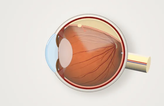
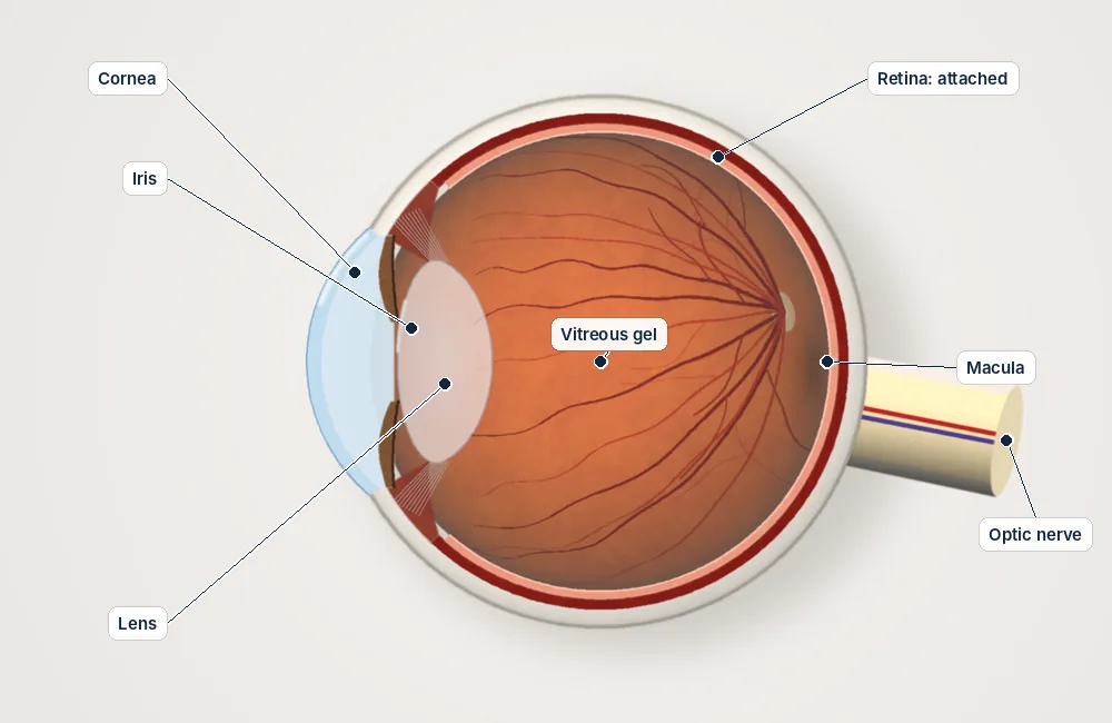
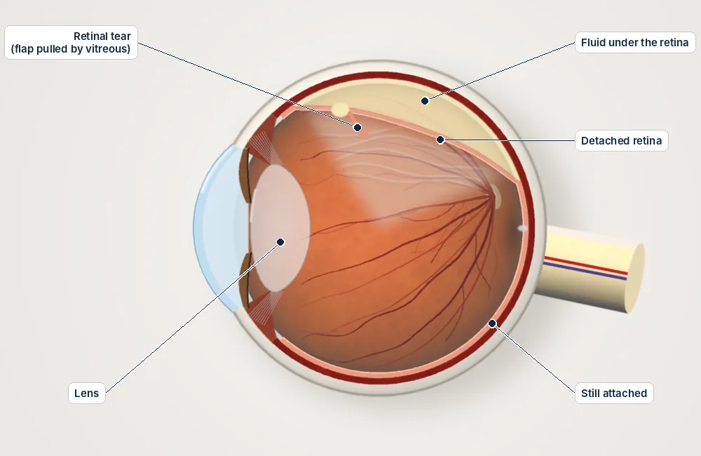
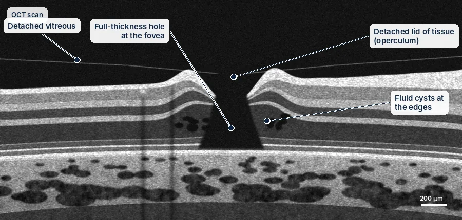
Inside the eye
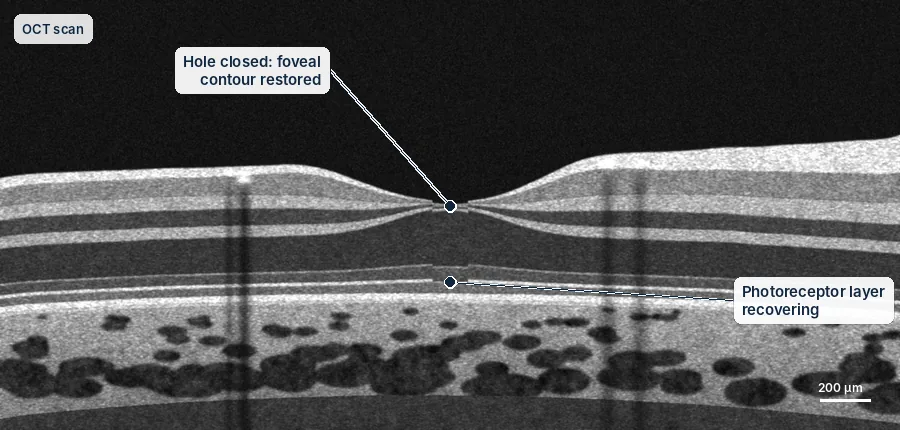
Vitrectomy repairs problems at the back of the eye from the inside, through tiny ports.
Simplified diagrams for patient education, not to scale.
Understanding the risks
Every procedure carries some risk. These are approximate rates from published studies, grouped by how often they happen. They are averages, not a prediction for you; Dr Weaver will discuss your own risks before any treatment.
Aftercare guides| Risk | Approximate rate |
|---|---|
| Common, usually temporary or manageable | |
| Cataract developing after vitrectomy | Most people over 50, within 1–2 years |
| Raised eye pressure | Common, usually temporary and treated with drops |
| Uncommon | |
| Retinal tear or detachment | About 1–2 in 100 |
| Further surgery needed | Depends on the condition; about 10–15 in 100 after retinal detachment repair |
| Rare but serious | |
| Infection inside the eye (endophthalmitis) | About 1 in 2,000–3,000 |
Everything you need to know
Tap a topic to open it.
Overview
A vitrectomy is delicate surgery inside the eye. The clear gel that fills the eye (the vitreous) is removed, so the surgeon can work directly on the retina and macula with very fine instruments. It is done through three tiny openings, each less than 1 mm wide, which usually seal on their own. Most vitrectomies are day surgery.
Conditions treated
- Retinal detachment
- Macular hole
- Epiretinal membrane and vitreomacular traction
- Bleeding inside the eye, including from diabetic eye disease
- Floaters that persist and really affect daily life (some cases)
- Lens implants that have moved, and pieces of lens left in the eye
- Eye injuries, including fragments inside the eye
How the surgery is done
- Numbing: local anaesthetic around the eye, with sedation so you are comfortable and relaxed. A general anaesthetic is used occasionally.
- Openings: three tiny openings are made in the white of the eye.
- Removing the gel: the gel is gently removed with a fine cutter, while fluid keeps the eye in shape.
- Repair: depending on the problem, scar tissue or membranes are peeled, the retina is flattened and laser seals any tears.
- Gas or oil: the eye may be filled with a gas bubble or silicone oil to hold the retina in place while it heals.
Surgery usually takes 30 to 90 minutes, depending on what is needed.
Gas bubbles and silicone oil
A gas bubble disappears on its own. SF6 gas usually takes about 2 weeks; longer-acting gases take 6 to 8 weeks. While there is gas in your eye, you must not fly, travel to high places or have nitrous oxide (laughing gas). You will wear a wristband until the gas has gone.
Silicone oil is used for some complex detachments. It is usually removed in a second, shorter operation after a few months.
Head positioning
If a gas bubble is used, holding your head in a set position helps the bubble press on the right place. For macular holes, Dr Weaver usually asks you to keep your face down for 45 minutes of every waking hour for at least 3 days. For a retinal detachment, the position depends on where the tear is. You will be given clear written instructions.
Recovery
- A check the day after surgery, then further visits over the following weeks
- Eye drops for about 4 weeks, and an eye shield at night for the first week
- Avoid heavy lifting, hard exercise and swimming for about 2 weeks
- Vision is blurred at first, especially with gas, and improves as the eye heals
- In people over 50, a vitrectomy usually makes a cataract form sooner, and cataract surgery is often needed within 1 to 2 years
You may also want to read
Talk to Dr Weaver about your eyes
A referral from your GP or optometrist is needed to claim Medicare rebates. Our team can help with bookings at Robina and Tweed Heads.