
Macular holeGold Coast & Tweed Heads
A small hole in the centre of the retina. It blurs and distorts central vision, making reading and faces hard to see. Surgery closes the hole in most people.
- Vitrectomy with SF6 gas
- Face-down positioning, usually at least 3 days
- Day surgery
The short version
- What is it?
- A small hole in the centre of the retina that blurs and distorts central vision.
- Will I lose my sight?
- A macular hole affects central vision only. Side vision is not affected.
- Can anything be done?
- Yes. Surgery closes the hole in about 90 in 100 typical cases.
- What does treatment involve?
- Day surgery (vitrectomy) with a gas bubble, then keeping your face down for most of each hour for at least 3 days. No flying until the gas has gone.
- What if I do nothing?
- Most holes stay open and central vision gradually gets worse. Small early holes sometimes close on their own, so they may be watched first.
The key facts
What you may notice
- Blurred or distorted central vision
- A missing spot in the centre of what you look at
- Straight lines that look bent
How it is diagnosed
- OCT scan confirms the size and stage
- Dilated examination of the macula
- Vision and distortion testing
How it is treated
- Vitrectomy removes the vitreous gel
- The innermost membrane is peeled to relax the edges of the hole
- An SF6 gas bubble supports the hole while it closes
Recovery
- Face-down 45 minutes of every waking hour, for at least 3 days
- No flying or high altitude until the gas has absorbed
- Reading vision recovers over weeks to months

Dr Travers Weaver
Macular hole surgery is part of Dr Weaver's subspecialty training in retinal surgery. He uses SF6 gas with 3 days of face-down positioning for most holes, and sees you the day after surgery.
- FRANZCOSpecialist eye surgeon
- 2 extra yearsRetinal surgery training, Vancouver
- About 1,000Operations a year
Inside the eye
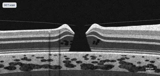
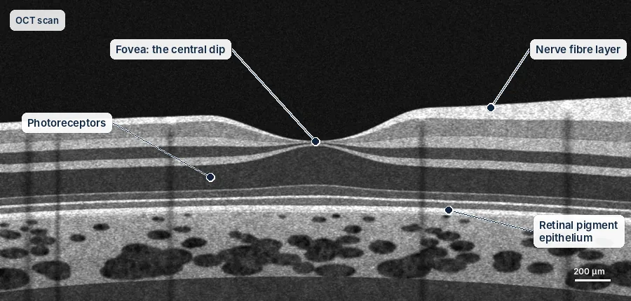
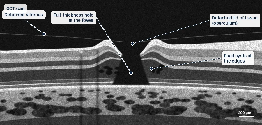
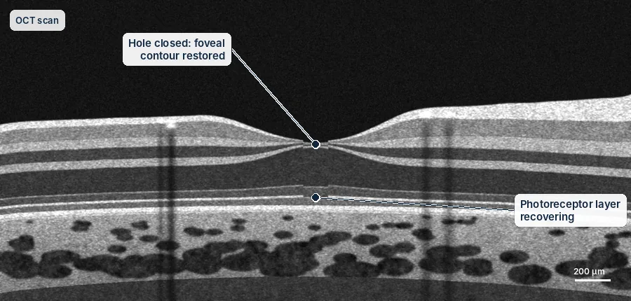
OCT scans show the retina in cross-section — the same view you will see on the screen in clinic.
Simplified diagrams for patient education, not to scale.
Understanding the risks
Every procedure carries some risk. These are approximate rates from published studies, grouped by how often they happen. They are averages, not a prediction for you; Dr Weaver will discuss your own risks before any treatment.
Aftercare guides| Risk | Approximate rate |
|---|---|
| Chance of success | |
| Hole closed with one operation | About 90 in 100 |
| Common, usually temporary or manageable | |
| Cataract developing after vitrectomy | Most people over 50, within 1–2 years |
| Raised eye pressure | Common, usually temporary and treated with drops |
| Uncommon | |
| Retinal tear or detachment | About 1–2 in 100 |
| Rare but serious | |
| Infection inside the eye (endophthalmitis) | About 1 in 2,000–3,000 |
Everything you need to know
Tap a topic to open it.
What is a macular hole?
A macular hole is a small opening in the centre of the retina, the part you use for reading and seeing faces. It causes blurred or distorted central vision, and sometimes a missing spot in the middle of what you look at. Surgery can close the hole in most people.
The macula is a small area at the centre of the retina. It gives you sharp, detailed central vision for reading, recognising faces and driving. A macular hole goes right through it.
Why holes form, and the stages
Most macular holes happen as part of normal ageing. The clear gel inside the eye (the vitreous) shrinks and peels away from the back of the eye. If the gel stays stuck to the macula as it peels away, it can pull a hole open.
Holes are graded from stage 1 to stage 4, by size and by how much the gel is still attached:
Stage 1
A hole is starting to form. It may settle on its own, so it is often just watched.
Stage 2
A small hole all the way through. Surgery is usually recommended.
Stage 3
A larger hole, with the gel still partly attached. Surgery is strongly recommended.
Stage 4
A larger hole, with the gel fully separated. Surgery is usually recommended.
The operation
The operation is done as day surgery. The eye is numbed and you are given sedation to relax you.
Removing the gel (vitrectomy)
Dr Weaver makes three tiny openings in the white of the eye, each less than 1 mm. He removes the gel so it can no longer pull on the macula.
Peeling the inner layer
This is the most important step. A very thin, clear layer on the surface of the retina (the internal limiting membrane) is stained with a dye so it can be seen. It is then gently peeled away with very fine instruments. This releases the forces that hold the hole open, and makes it much less likely to come back.
The gas bubble
The eye is filled with a gas bubble (SF6). The bubble floats up and presses gently on the macula, holding the edges of the hole together while it heals. The gas slowly disappears over about 2 weeks.
No flying while there is gas in your eye
You must not fly or travel to high places while gas is in your eye. The gas expands at altitude and can cause a dangerous rise in eye pressure, with severe pain and loss of vision. You will wear a wristband until the gas has gone. Keep it on, and show it to any doctor, dentist or anaesthetist who treats you. Nitrous oxide (laughing gas), used in some anaesthetics, must not be given while gas is in your eye.
Face-down positioning after surgery
Keeping your face down after surgery is one of the most important things you can do to help the hole close. The gas bubble floats upwards. The macula is at the back of the eye, so to make the bubble press on it, you need to face the floor.
The position
Look at the floor, not the ceiling. You can sit with your chin on your chest, or lie face-down. A posturing pillow, or a hired face-down support such as a massage-style face rest or a mirror, makes it easier. Dr Weaver's team will give you details before surgery.
The usual plan:
- How long: face-down for 45 minutes of every waking hour, for at least 3 days after surgery. Dr Weaver may ask you to keep going for longer, depending on the size of the hole.
- Sleep: face-down or on your side. Do not sleep on your back.
- Breaks: use the other 15 minutes of each hour to eat, drink and go to the bathroom. A posturing mirror lets you read or watch television.
- Help at home: arrange a lift home, and someone to help with meals and errands for the first few days, as you will be positioning most of the day and cannot drive.
- Larger holes (stage 3 or 4) may need a longer period. Dr Weaver will advise you, based on your scan and what he finds during surgery.
Equipment
Face-down equipment makes this much more comfortable. Options include pillows with a face hole, face cradles that fit a table or chair, mirrors so you can watch television while face-down, and supports for overnight. The practice will give you supplier contacts before your surgery date.
Gas bubble safety rules
- No flying until the gas has gone, which takes about 2 weeks. Check with Dr Weaver before booking any travel.
- No nitrous oxide (laughing gas). Tell any anaesthetist that you have gas in your eye. Nitrous oxide can enter the bubble and cause a dangerous rise in eye pressure.
- Wear your gas wristband until the gas has gone.
- No scuba diving until the gas has gone.
- Avoid high places: do not travel above about 1,000 metres, for example over mountain roads.
- No driving while there is gas in your eye, as your vision will be very blurred. You will need someone to drive you home and to take you places until the gas has gone.
Results and recovery
Published studies report that about 90 in 100 typical macular holes close after one operation. Larger holes, and holes that have been there a long time, are less likely to close. For larger holes, Dr Weaver may use extra techniques, such as a wider peel or a small flap of the inner layer placed over the hole.
Closing the hole and seeing well are not the same thing.
- Closing the hole: happens in about 90 in 100 typical holes.
- Sharper vision: improves in most people, but by different amounts.
- Less distortion: most people notice straight lines look straighter.
- Some blur or distortion may remain, especially if the hole was large or had been there a long time.
Vision usually starts to improve 4 to 6 weeks after surgery and keeps improving slowly for up to 12 months. How much it improves depends on the size of the hole, how long it has been there, and any damage before surgery. Most people notice less distortion early on, and sharper vision comes back more gradually.
For holes that have been there more than a year, or that did not close after an earlier operation, other surgical techniques may be considered.
You may also want to read
Talk to Dr Weaver about your eyes
A referral from your GP or optometrist is needed to claim Medicare rebates. Our team can help with bookings at Robina and Tweed Heads.