
Retinal detachmentGold Coast & Tweed Heads
The retina lifts away from the back of the eye. It is an emergency, and in many cases it can be repaired with surgery.
- Urgent assessment
- Vitrectomy or scleral buckle
- Day surgery
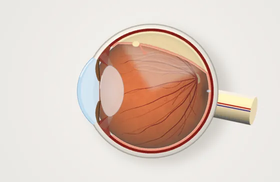
Symptoms can be urgent. New floaters, flashes, a shadow or sudden vision loss need same-day assessment. Call (07) 5612 5200, or after hours go to the emergency department at Gold Coast University Hospital.
The short version
- What is it?
- The light-sensing layer at the back of the eye comes away from the wall of the eye, usually starting from a tear.
- Will I lose my sight?
- Without surgery, vision in that eye is usually lost. Prompt repair gives the best chance of good vision.
- Can anything be done?
- Yes. Surgery reattaches the retina with one operation in about 85 to 90 in 100 people.
- What does treatment involve?
- Day surgery, usually a vitrectomy with a gas bubble, then holding a head position for several days. No flying with gas in the eye.
- What if I do nothing?
- A detachment almost always spreads. This is an emergency: call the rooms the same day, or after hours go to Gold Coast University Hospital emergency.
The key facts
What you may notice
- A shower of new floaters
- Flashes of light at the edge of vision
- A shadow or curtain spreading across vision
How it is diagnosed
- Dilated examination of the whole retina
- Wide-field retinal photography
- Ultrasound if blood or cataract blocks the view
How it is treated
- Vitrectomy: the gel is removed and the retina flattened
- Scleral buckle: a band on the outside of the eye supports the tear
- Laser or freezing treatment seals the tear
- A gas bubble or silicone oil supports the retina while it heals
Recovery
- Head posturing for several days with gas or oil
- Vision is very blurred through a gas bubble until it dissolves
- No flying or high altitude while gas is in the eye

Dr Travers Weaver
Repairing retinal detachments is a core part of Dr Weaver's subspecialty training and his everyday work. He performs vitrectomy and scleral buckle surgery, and sees you the day after a vitrectomy.
- FRANZCOSpecialist eye surgeon
- 2 extra yearsRetinal surgery training, Vancouver
- About 1,000Operations a year
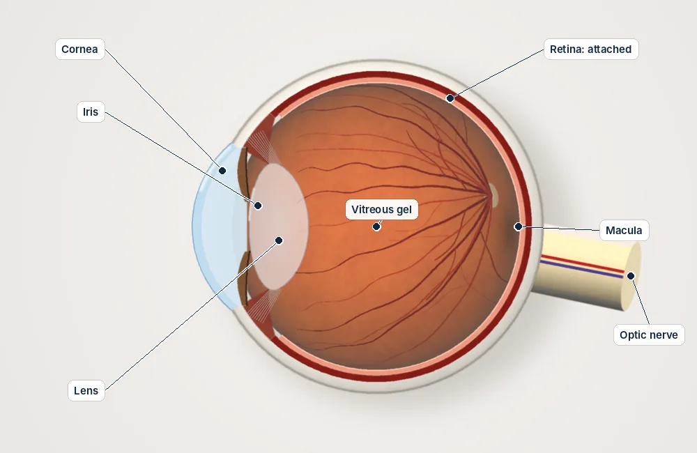
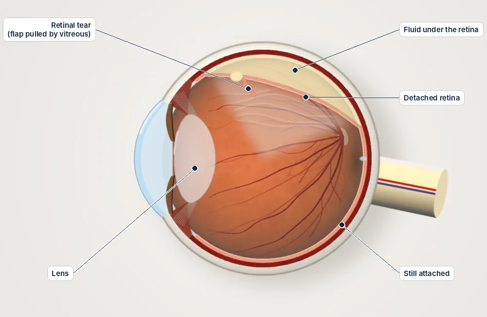
Inside the eye
A tear lets fluid pass under the retina and lift it away from the back wall of the eye.
Simplified diagrams for patient education, not to scale.
Understanding the risks
Every procedure carries some risk. These are approximate rates from published studies, grouped by how often they happen. They are averages, not a prediction for you; Dr Weaver will discuss your own risks before any treatment.
Aftercare guides| Risk | Approximate rate |
|---|---|
| Chance of success | |
| Retina reattached with one operation | About 85–90 in 100 |
| Common, usually temporary or manageable | |
| Cataract developing after vitrectomy | Most people over 50, within 1–2 years |
| Raised eye pressure | Common, usually temporary and treated with drops |
| Scleral buckle: change in glasses prescription | Common; usually more short-sighted, new glasses once the eye settles |
| Uncommon | |
| Re-detachment needing further surgery | About 10–15 in 100 |
| Scleral buckle: double vision | Uncommon, usually temporary |
| Rare but serious | |
| Infection inside the eye (endophthalmitis) | About 1 in 2,000–3,000 |
| Scleral buckle: buckle exposure or infection | Rare; the buckle may need to be removed |
Everything you need to know
Tap a topic to open it.
What is a retinal detachment?
The retina is the thin, light-sensing layer that lines the back of the eye, like the film in a camera. A retinal detachment is when the retina comes away from the wall of the eye, which supplies it with oxygen and nourishment. Without surgery, the cells of the retina start to die within hours to days, and vision is lost for good. It is a true eye emergency.
The most common type starts with a tear in the retina. Fluid from inside the eye passes through the tear and gradually lifts the retina away, like wallpaper peeling off a damp wall. Less often, a detachment is caused by scar tissue pulling on the retina, or by fluid building up without a tear.
Warning signs: act straight away
Never ignore these symptoms. Call the rooms straight away on (07) 5612 5200 during business hours. After hours, go to the emergency department at Gold Coast University Hospital.
- A sudden shower of new floaters: dark spots, cobwebs, threads or rings drifting across your vision
- Flashes of light, especially at the edge of your vision and in dim light
- A dark curtain, shadow or veil over part of your vision. This means the detachment is spreading.
- Sudden blurring or loss of central vision. This suggests the centre of the retina (the macula) has come away.
Time matters. If the detachment is repaired before it reaches the macula, the chance of good vision is much higher than if the macula has already come away.
Who is at higher risk?
- Short-sightedness (myopia), especially –6 or stronger. The retina is thinner and more stretched.
- A previous retinal detachment in either eye
- A family history of retinal detachment
- Previous cataract surgery, especially if there were complications
- An injury to the eye
- Lattice degeneration, which is thin, weak areas at the edge of the retina
- A recent posterior vitreous detachment, when the gel inside the eye separates from the retina, especially if it caused a tear
The operation: vitrectomy
For most detachments, Dr Weaver repairs the retina with an operation called a vitrectomy. It is usually done as day surgery with the eye numbed and sedation to relax you. A general anaesthetic is used when needed.
Getting ready
Local anaesthetic is injected around the eye to numb it, and you are given sedation through a drip so you are comfortable and relaxed. The eye and skin around it are cleaned with antiseptic.
Removing the gel
Dr Weaver makes three tiny openings, each less than 1 mm, in the white of the eye. Through these he uses a fine light, a small fluid line and a cutting instrument to remove the gel that is pulling on the retina. He works under an operating microscope.
Finding and treating the tear
Using a wide-angle viewing lens, he finds every tear or hole in the retina. The fluid under the retina is drained, or pushed out using a heavy liquid that gently flattens the retina from the inside.
Sealing the tear
Once the retina is flat, laser (or, rarely, freezing treatment) is applied around each tear. This forms a scar that seals the retina down permanently, so fluid cannot get back underneath.
Gas or oil
The eye is filled with a gas bubble or silicone oil, which holds the retina in place while it heals. Dr Weaver chooses the best option for your detachment.
Gas bubble or silicone oil?
After a vitrectomy, the eye is filled with gas or oil to press the retina against the wall of the eye while it heals. Dr Weaver will explain which is best for you.
Gas bubble (SF6 or C3F8)
A gas bubble is used most often. SF6 gas disappears over about 2 weeks. C3F8 lasts longer, 6 to 8 weeks. You must not fly until the gas has gone. You will usually need to hold a set head position for several days.
Silicone oil
Oil is used for more complex detachments, such as those with scar tissue, very large tears or tears low down in the eye. The oil stays in the eye until a second, smaller operation removes it, usually after 3 to 6 months. You still need to hold a head position after surgery so the oil supports the repair. Flying is generally allowed with oil in the eye.
No flying with gas in your eye
You must NOT fly or travel to high places while you have a gas bubble in your eye. The gas expands at altitude and can cause a dangerous, painful rise in eye pressure. Tell any doctor, dentist or anaesthetist that you have gas in your eye, because nitrous oxide (laughing gas) must not be used. You will wear a wristband until the gas has gone, so anyone treating you knows.
Head positioning after surgery
Holding your head in a set position after surgery (called posturing) is one of the most important things you can do to help the repair succeed. The gas or oil needs to press against the sealed tear while the laser scar becomes strong, so fluid cannot get back under the retina.
Why it works
Gas and oil float. Positioning your head so the bubble floats up against the tear keeps steady, gentle pressure on the repair. Even short breaks can let fluid seep back under the retina before the scar has formed.
Your position depends on where the tear is:
- Tears in the upper retina: sit or stand with your head upright, so the bubble floats up against the repair.
- Tears at the side: lie on the opposite side, so the affected side is uppermost.
- Tears in the lower retina or at the macula: you may need to keep your face down or your head tilted, or silicone oil may be used instead.
Tips
Positioning cushions and face-down supports, such as face cradles and mirrors for watching television, can be hired or bought. Dr Weaver's team will tell you how to get them before surgery. Dr Weaver will tell you which position to hold and for how long, which is usually most of the waking day for the first few days. The more closely you keep to it, the better your chance of success. You can eat, drink and use the bathroom as normal; just keep the position as much as you can. Most people hold the position closely for the first 5 days.
Scleral buckle
Sometimes Dr Weaver recommends a scleral buckle instead of, or as well as, a vitrectomy. This is more likely for younger people whose gel has not yet separated from the retina, or for some detachments low in the eye. A soft silicone band is stitched to the outside of the eye. It gently pushes the wall of the eye inwards, against the tear, and relieves the pulling on the retina. A gas bubble is not usually needed.
Recovery after a scleral buckle: the operation is done with the eye numbed and sedation, or under a general anaesthetic, usually as day surgery. For the first 1 to 2 weeks the eye is often more swollen, red and achy than after a vitrectomy, and regular pain relief helps. The band stays in place for good and cannot be seen. It slightly changes the shape of the eye and usually makes it more short-sighted, so you will need new glasses once the eye has settled.
Risks of a scleral buckle include a change in glasses prescription, double vision (usually temporary), raised eye pressure and, rarely, the band showing through or becoming infected, which may mean it needs to be removed. As with any retinal detachment surgery, the retina can come away again and further surgery may be needed.
Recovery and results
How well you see after surgery depends mostly on whether the macula had come away before surgery. If the macula was still attached, vision often recovers well, sometimes close to what it was before. If the macula had come away, you can still get back useful vision, but central vision may stay somewhat reduced, even when the retina is successfully reattached.
If a gas bubble is used, vision is very blurred through the bubble at first. As the gas slowly dissolves, you will see a line across your vision that moves lower each day until the bubble has gone. Most people then notice their vision slowly improve over weeks to months. The eye may be red and achy for the first 1 to 2 weeks. Your glasses prescription often changes, so have it checked once the eye has fully settled, usually 2 to 3 months after surgery.
Retinal detachment: common questions
Is a retinal detachment an emergency?
Yes. A retinal detachment needs urgent assessment and usually surgery within days, sooner if central vision is still good. If you notice a shower of new floaters, flashes or a shadow spreading across your vision, call the rooms on (07) 5612 5200 the same day.
Where should I go if I think I have a retinal detachment?
During business hours, call the rooms on (07) 5612 5200 so you can be seen urgently. After hours, go to the emergency department at Gold Coast University Hospital. Do not wait for a routine appointment.
How successful is retinal detachment surgery?
Published studies report that about 85–90 in 100 detachments are repaired with one operation, and most of the rest with further surgery. How well you see afterwards depends mostly on whether the centre of the retina (the macula) was detached.
How long until I can fly after retinal detachment surgery?
You must not fly while gas is in the eye. SF6 gas usually lasts about 2 weeks and longer-acting gases 6–8 weeks. Flying is usually possible with silicone oil. Dr Weaver will confirm when the gas has gone.
What is the recovery like?
Most people go home the same day. You may need to hold a particular head position for several days, use drops for about 4 weeks and avoid heavy lifting. If a gas bubble is used, vision is very blurred through the bubble at first. As the gas gradually dissolves you will see a line across your vision that moves lower each day until the bubble has gone. Vision then continues to improve over weeks to months.
Can it happen in my other eye?
The risk in the other eye is higher than average, particularly if you are short-sighted. Know the warning signs and have the other eye checked as advised.
You may also want to read
Talk to Dr Weaver about your eyes
A referral from your GP or optometrist is needed to claim Medicare rebates. Our team can help with bookings at Robina and Tweed Heads.