
Eye tests explained
What the scans and measurements at your appointment do, and how to check your own central vision at home.
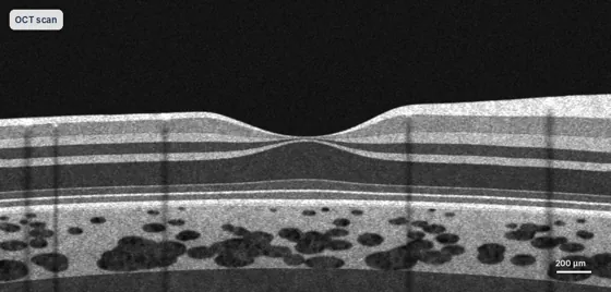
Optical coherence tomography (OCT)
A non-contact scan that shows the layers of the retina in cross-section, like an ultrasound that uses light. It detects fluid, holes, membranes and thinning, and guides injection treatment.
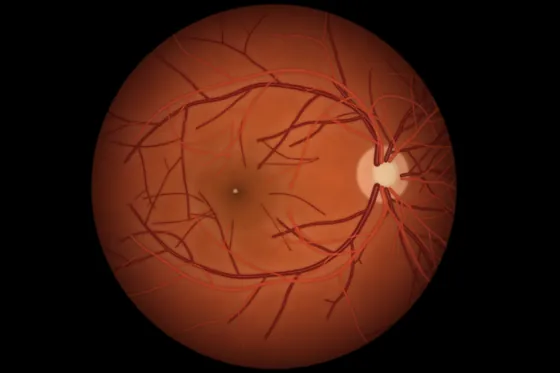
Wide-field retinal photography
High-resolution photographs of the retina, out to the periphery, usually taken through a dilated pupil. They record changes such as freckles, bleeding or new vessels so they can be compared over time.
Biometry (IOLMaster)
A precise optical measurement of the length of the eye and the curvature of the cornea, used to calculate the power of the lens implant for cataract surgery and refractive lens exchange.
Corneal tomography (Pentacam)
A rotating camera maps the front and back surfaces and thickness of the cornea, measuring astigmatism and helping plan lens choice, including toric lenses.
Wavefront and topography (Nidek OPD)
Measures how light is focused through the cornea and lens, including subtle optical irregularities and pupil size. It helps when choosing a lens implant and when investigating unexpected blur or glare.
Specular microscopy
Photographs the inner cell layer of the cornea (the endothelium) and counts the cells. It is important before cataract or lens surgery in eyes with corneal conditions such as Fuchs dystrophy.
B-scan ultrasound
A small probe on the closed eyelid uses sound waves to image the back of the eye when blood or a dense cataract blocks the view. It also measures the thickness of tumours.
Fluorescein angiography (FFA)
A dye is injected into a vein in the arm and photographs track it through the retinal vessels, showing leaks and blockages. The dye can turn the skin and urine yellow for a day. Tell us about any allergies.
Visual field testing
You press a button when you see small lights in your side vision. It maps the field of vision and is important for glaucoma, some retinal conditions and driving or CASA assessments.
Amsler grid
A quick daily check for people with macular degeneration, macular holes or other macular conditions.
- Hold the screen or printout at reading distance in good light, wearing your reading glasses.
- Cover one eye and look steadily at the central dot.
- Check that all the lines are straight and every square is present, then repeat with the other eye.
Wavy, broken or missing lines? Call the rooms promptly on (07) 5612 5200. New distortion can be a sign of wet macular degeneration, which is best treated early.
Talk to Dr Weaver about your eyes
A referral from your GP or optometrist is needed to claim Medicare rebates. Our team can help with bookings at Robina and Tweed Heads.