
Vitreomacular tractionGold Coast & Tweed Heads
The vitreous gel stays stuck to the macula and pulls on it, distorting vision.
- OCT diagnosis
- Often monitored
- Vitrectomy if needed
The key facts
What you may notice
- Distortion of straight lines
- Blurred central vision
- Sometimes no symptoms at all
How it is diagnosed
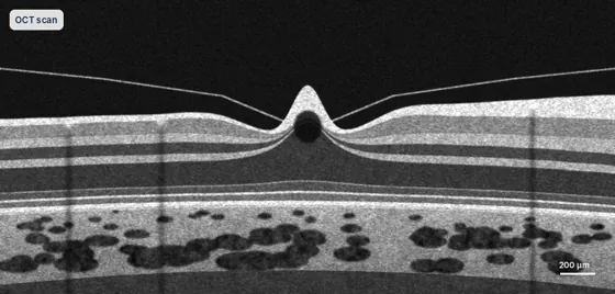
- OCT scan shows the gel pulling on the macula
- Dilated examination
- Amsler grid to monitor distortion
How it is treated
- Can release on its own, but not often; monitored with OCT
- Vitrectomy to release the traction if vision is affected
- Treatment of any macular hole that develops
Recovery
- Regular OCT scans
- Check each eye at home with an Amsler grid
- Report any new distortion promptly

Dr Travers Weaver
Dr Weaver monitors traction with OCT scans in his rooms and performs the surgery himself if it is needed, seeing you the day after a vitrectomy.
- FRANZCOSpecialist eye surgeon
- 2 extra yearsRetinal surgery training, Vancouver
- About 1,000Operations a year
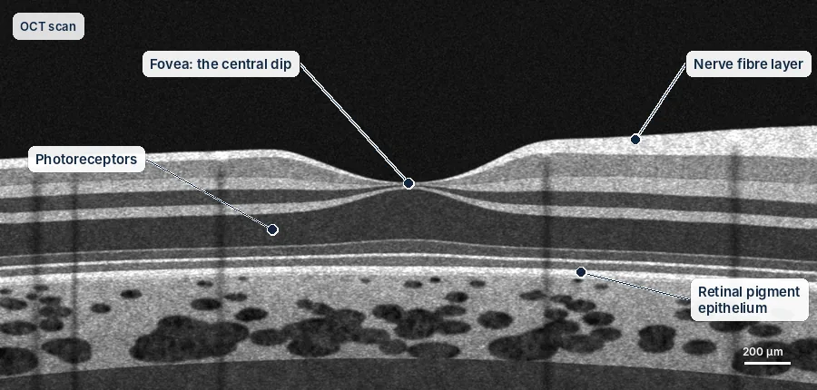
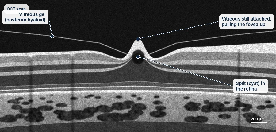
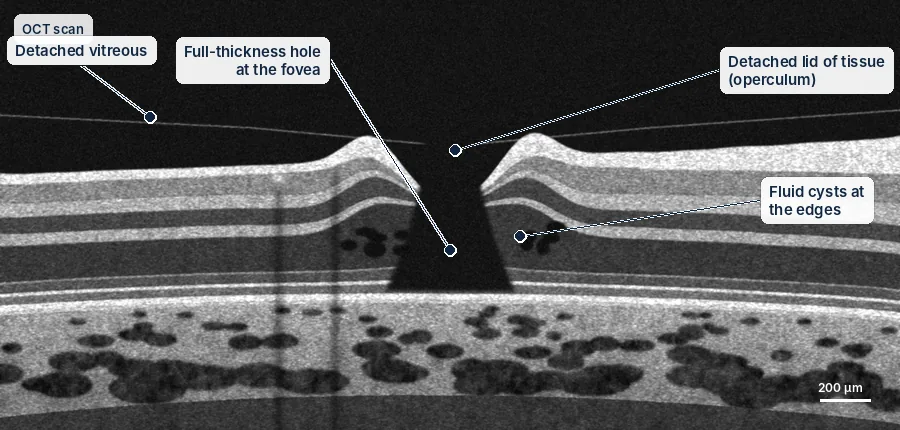
Inside the eye
Persistent pulling by the vitreous can distort the fovea and, in some eyes, progress to a macular hole.
Simplified diagrams for patient education, not to scale.
Understanding the risks
Every procedure carries some risk. These are approximate rates from published studies, grouped by how often they happen. They are averages, not a prediction for you; Dr Weaver will discuss your own risks before any treatment.
Aftercare guides| Risk | Approximate rate |
|---|---|
| Common, usually temporary or manageable | |
| Cataract developing after vitrectomy | Most people over 50, within 1–2 years |
| Raised eye pressure | Common, usually temporary and treated with drops |
| Uncommon | |
| Retinal tear or detachment | About 1–2 in 100 |
| Rare but serious | |
| Infection inside the eye (endophthalmitis) | About 1 in 2,000–3,000 |
Everything you need to know
Tap a topic to open it.
Overview
Dr Weaver sees people with vitreomacular traction at his rooms in Robina and Tweed Heads. An OCT scan shows how strongly the gel is pulling, and this guides what to do.
What is vitreomacular traction?
The inside of the eye is filled with a clear gel called the vitreous. With age, this gel normally shrinks and peels away from the back of the eye. This is called a posterior vitreous detachment (PVD).
Sometimes the gel does not come away cleanly. It stays stuck to the very centre of the retina (the macula) and pulls on it. This is vitreomacular traction (VMT). The pulling changes the shape of the macula, so central vision becomes blurred or distorted.
VMT is part of a range. At the mild end, the gel is stuck on but not pulling (vitreomacular adhesion). At the severe end, the pulling tears a hole in the macula (a macular hole).
The stages
- Gel attached, not pulling (vitreomacular adhesion): the retina keeps its normal shape. There are usually no symptoms, and checks are all that is needed.
- Gel pulling (vitreomacular traction): the scan shows the centre of the retina being pulled out of shape. This can affect vision and can lead to a macular hole.
- Pulling with a partial hole (lamellar hole): the pulling has opened a hole part of the way through the macula. This needs careful monitoring.
Symptoms
- Blurred central vision
- Straight lines look wavy or bent (metamorphopsia)
- Things look smaller than they should (micropsia)
- Some people notice very little, even when the scan shows clear changes
Treatment options
Watching and waiting. The gel can let go on its own, but this does not happen often. If you have few or no symptoms and your scans are stable, regular checks with an OCT scan every 3 to 6 months are a sensible first step.
Vitrectomy surgery. Surgery is the most reliable treatment if VMT is causing symptoms, is getting worse, or has caused a hole. The gel is removed so it can no longer pull. If there is a hole, a very thin layer on the surface of the retina is also peeled away to help it close. Surgery relieves the pulling in most people, and most notice their vision improve.
Could it become a macular hole?
Yes. People with VMT are more likely to develop a full macular hole. The risk is higher when the gel is stuck to a small area, or when scans show the centre of the retina changing over time. Dr Weaver looks for these signs at each visit and will suggest surgery earlier if things are getting worse.
You may also want to read
Talk to Dr Weaver about your eyes
A referral from your GP or optometrist is needed to claim Medicare rebates. Our team can help with bookings at Robina and Tweed Heads.