
Ocular oncologyGold Coast & Tweed Heads
Assessment and monitoring of freckles (naevi) and other growths inside the eye.
- Naevus monitoring
- Detailed imaging
- Coordinated care
The key facts
What you may notice
- Often found on a routine eye test
- Sometimes blurred vision or flashes
- A shadow in part of your vision
How it is diagnosed
- Wide-field photography to record size and colour
- OCT and B-scan ultrasound to measure thickness
- Autofluorescence imaging
How it is treated
- Imaging with photography, OCT and ultrasound
- Monitoring benign naevi for any change
- Referral for specialised treatment when needed
Recovery
- Review intervals are set by risk
- Bring any previous eye photos with you
- Report new symptoms promptly
Inside the eye
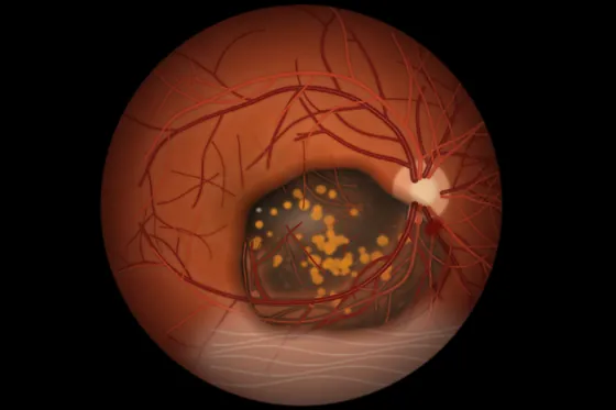
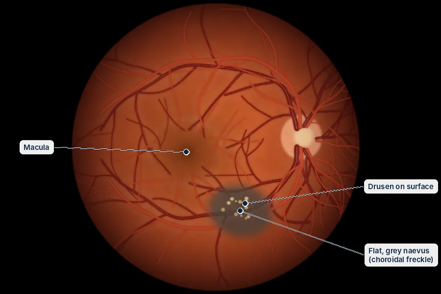
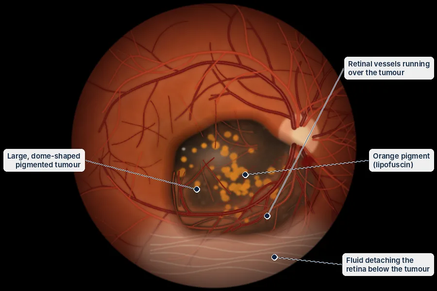
Most pigmented spots at the back of the eye are benign naevi; features such as thickness, fluid and orange pigment warrant closer review.
Simplified diagrams for patient education, not to scale.
Everything you need to know
Tap a topic to open it.
Overview
Dr Weaver assesses and monitors tumours inside the eye at his rooms in Robina and Tweed Heads. Complex cases are managed together with cancer specialists, radiation oncologists and pathologists.
Tumours of the eye
Tumours can grow from different tissues inside the eye. Many are harmless and only need to be watched. Others can threaten sight, or life, and need treatment. Finding them early, often at a routine eye check, makes a big difference. Dr Weaver assesses and manages a wide range of tumours at the back of the eye.
Choroidal melanoma
Choroidal melanoma is the most common cancer that starts inside the eye in adults. It grows from pigment cells in the layer under the retina (the choroid). It may be found by chance at a routine eye check, or cause blurred vision, flashes, floaters or a missing patch of vision. Treatment depends on the size and position of the tumour:
- Close monitoring: small tumours with low-risk features may be watched closely with regular scans and photographs.
- Plaque radiotherapy (brachytherapy): the standard treatment for medium-sized melanomas. A small radioactive disc is stitched to the outside of the eye over the tumour for a few days.
- Removal of the eye: needed for very large tumours.
- Proton beam radiotherapy: available at some centres for some tumours.
Everyone with a choroidal melanoma needs scans of the body and ongoing checks to make sure it has not spread. This is coordinated with a cancer specialist.
Choroidal naevus (a freckle in the eye)
A choroidal naevus is a freckle at the back of the eye. About 5 to 10 in every 100 people have one. Almost all are harmless and only need checking from time to time. A few have features that mean they need closer checks, or referral for treatment:
- Thicker than 2 mm
- Fluid under the retina over the freckle
- Changes in vision
- Orange pigment on its surface
- Its edge is close to the optic nerve
- No small yellow deposits (drusen) on it
- It looks hollow on ultrasound
Other tumours of the eye
- Retinal haemangioma: a tumour made of blood vessels in the retina. It may occur on its own or as part of Von Hippel-Lindau syndrome. It is treated with laser, eye injections or freezing treatment.
- Choroidal haemangioma: a harmless tumour made of blood vessels under the retina. If it affects central vision, it is treated with a light-activated treatment (photodynamic therapy).
- Retinoblastoma: the most common eye tumour in children. It needs urgent referral to a specialist children's team.
- Cancer that has spread to the eye: the layer under the retina is the most common place in the eye for cancer to spread to, most often from the breast, lung or bowel. Care is coordinated with your cancer specialist.
- Lymphoma: lymphoma inside the eye may cause floaters, inflammation and patches on the retina. A small sample of the gel (a vitreous biopsy) is often needed to confirm it.
You may also want to read
Talk to Dr Weaver about your eyes
A referral from your GP or optometrist is needed to claim Medicare rebates. Our team can help with bookings at Robina and Tweed Heads.