
Epiretinal membraneGold Coast & Tweed Heads
A thin layer of scar tissue on the surface of the macula that wrinkles the retina and distorts vision.
- Vitrectomy and membrane peel
- Usually no posturing
- Day surgery
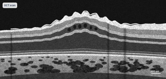
The key facts
What you may notice
- Straight lines look wavy
- Blurred central vision
- Objects look a different size in each eye
How it is diagnosed
- OCT scan shows the membrane
- Dilated examination
- Distortion testing with an Amsler grid
How it is treated
- Observation if symptoms are mild
- Vitrectomy with peeling of the membrane
- Often a small air bubble; face-down positioning routinely not needed
Recovery
- Distortion eases gradually over several months
- Eye drops for a few weeks
- Follow-up OCT scans track recovery

Dr Travers Weaver
Membrane peeling is part of Dr Weaver's subspecialty training in retinal surgery, including repeat operations. Face-down positioning is routinely not needed, and he sees you the day after surgery.
- FRANZCOSpecialist eye surgeon
- 2 extra yearsRetinal surgery training, Vancouver
- About 1,000Operations a year
Inside the eye
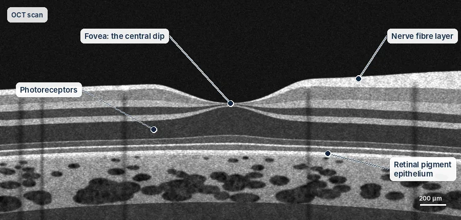
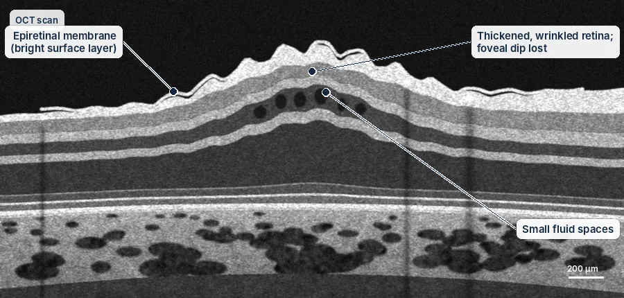
OCT scans show the retina in cross-section — the same view you will see on the screen in clinic.
Simplified diagrams for patient education, not to scale.
Understanding the risks
Every procedure carries some risk. These are approximate rates from published studies, grouped by how often they happen. They are averages, not a prediction for you; Dr Weaver will discuss your own risks before any treatment.
Aftercare guides| Risk | Approximate rate |
|---|---|
| Chance of success | |
| Distortion improves | Most people, gradually over months |
| Common, usually temporary or manageable | |
| Cataract developing after vitrectomy | Most people over 50, within 1–2 years |
| Raised eye pressure | Common, usually temporary and treated with drops |
| Uncommon | |
| Membrane returns | About 5 in 100 |
| Retinal tear or detachment | About 1–2 in 100 |
| Rare but serious | |
| Infection inside the eye (endophthalmitis) | About 1 in 2,000–3,000 |
Everything you need to know
Tap a topic to open it.
Overview
Dr Weaver performs surgery for epiretinal membranes, including complex cases and repeat operations. He consults at Robina and Tweed Heads and operates at Miami Private Hospital, Tweed Day Surgery and Pacific Private Hospital.
What is an epiretinal membrane?
An epiretinal membrane is a thin, see-through layer of scar tissue that grows on the surface of the macula, the centre of the retina that you use for reading and fine detail. It is also called a macular pucker. As the membrane tightens, it wrinkles the retina underneath, a bit like cling film shrinking on a surface. This blurs and distorts your vision.
Most membranes form after the gel inside the eye separates from the retina with age (a posterior vitreous detachment). They can also follow a retinal tear, retinal detachment surgery or inflammation. Often there is no clear cause.
Symptoms
- Straight lines look wavy or bent (metamorphopsia)
- Blurred central vision
- A ghost image or double vision in that eye
- Trouble reading, especially small print
- Things look smaller than they should (micropsia)
- Symptoms usually come on slowly, over months to years
An OCT scan, a quick and painless photo of the layers of the retina, shows the membrane and how much it is wrinkling the retina. This helps decide when surgery is worthwhile.
When is surgery recommended?
Not every membrane needs surgery. Mild membranes that do not bother you much can simply be checked with scans and vision tests. Surgery is considered when:
- The distortion is affecting your daily life
- Your vision has dropped a lot
- Scans show the membrane is getting worse
- It is getting in the way of driving, reading or work
Dr Weaver will look carefully at your symptoms, vision and scans before recommending surgery. The right time is different for each person.
The operation: vitrectomy and membrane peel
The operation is done as day surgery. The eye is numbed and you are given sedation to relax you. It usually takes 30 to 45 minutes.
- Vitrectomy: the gel inside the eye is removed through three tiny openings, each less than 1 mm, so the surface of the retina can be reached.
- Peeling the membrane: using very fine instruments and high magnification, the membrane is gently lifted off the retina. This needs great care so the retina underneath is not damaged.
- Peeling the inner layer: in most cases Dr Weaver also peels a very thin layer on the surface of the retina (the internal limiting membrane). A dye makes it visible. This lowers the chance of the membrane coming back.
- At the end: a small air bubble is often left in the eye to help the openings seal. It goes away within a few days. A long-lasting gas bubble is not usually needed, and you will not routinely need to keep your face down.
Recovery and what to expect
Vision improves slowly, over 3 to 12 months, as the retina flattens out and recovers. Most people notice less distortion and clearer vision. Vision does not always return completely to normal, especially if the membrane has been there for many years or has damaged the retina.
The aim of surgery is to stop things getting worse and to improve vision. It may not make your vision perfect. Dr Weaver will talk with you about what to expect, based on your own scans.
You will not routinely need to keep your face down. If a small air bubble was left in the eye, your vision will be blurred for a few days until it goes, and you must not fly until it has gone. Do not rub the eye, and avoid swimming and heavy lifting for 2 weeks.
You may also want to read
Talk to Dr Weaver about your eyes
A referral from your GP or optometrist is needed to claim Medicare rebates. Our team can help with bookings at Robina and Tweed Heads.